Ferrofluid Droplets to Locally Measure the Mechanics of Soft Materials
Brief Description
A technique and apparatus that can measure the mechanical properties of any kind of soft material, including complex fluids, living embryonic and adult tissues (such as skin), as well as tumors.
Background
Current methodologies to measure the mechanical (material) properties of soft materials do not allow local and/or spatiotemporal measurements of the material properties at different length scales. These limitations are particularly relevant for biological samples, from single cells to embryonic tissues and disease processes (such as cancer), which change in space and time. Also, in the case of soft materials, such as complex fluids (emulsions, foams, polymer melts, etc.), no single technique allows the measurement of their mechanics at different length scales as well as spatially and temporally. The technique described below overcomes all of these limitations.
Description
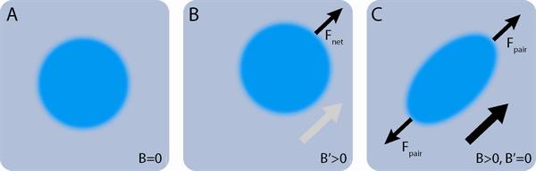
UC Santa Barbara researchers have developed a technique and apparatus that can measure the mechanical properties of any kind of soft material, including complex fluids, living embryonic and adult tissues (such as skin), as well as tumors. The apparatus applies controlled forces and measures mechanical properties of a large variety of samples by detecting the shape dynamics of a biocompatible ferrofluid drop in the presence of controlled magnetic fields (Figure 1). The technique allows for the measurement of mechanics along any spatial direction (mechanical anisotropy) and any length scale ranging from 1 micrometer to several millimeters. Also, it can perform simultaneous measurements at different locations of the sample, even if the sample is changing over time, providing a spatiotemporal map of the local mechanical properties of the material. For biological applications, it allows measurements at the subcellular, cellular and larger tissue scales, all in situ and in vivo. It is also compatible with fluorescent and other optical microscopy.
Figure 1: Scheme to apply forces using ferrofluid droplets in a magnetic field.
Otger Campus' Faculty Page: http://www.
Publication Link: http://www.nature.com/
Advantages
- First technique to measure local mechanical properties in living tissues
- Permits measurement of local material properties at different scales
- Permits measurement of local mechanical anisotropy
- Permits measurement of material properties changing in space and time
- Can be used in a broad array of samples including any soft material or polymer gel, and living materials such as tissues, both embryonic and adult
Applications
- Life sciences research
- Materials science research
- Cancer diagnostics
- Magnetic particle imaging
- Non-destructive mechanical measurements within 3D tissues
Embryonic stem cells
Tissue grafts (e.g. skin)
Cosmetic research
Diagnostics - Non-destructive mechanical measurements within 3D materials
Implants
Material science research
Patent Status
| Country | Type | Number | Dated | Case |
| United States Of America | Issued Patent | 10,149,630 | 12/11/2018 | 2015-300 |
Contact
- Donna M. Cyr
- cyr@tia.ucsb.edu
- tel: View Phone Number.

Inventors
- Campas, Otger
- Serwane, Friedhelm
Other Information
Keywords
ferrofluidic, magnetic forces, stem cell, diagnostics, indansens