Phasor method to fluorescence lifetime microscopy discriminates metabolic states of germ cells in a living tissue / Phase method to fluorescence lifetime microscopy to discriminate metabolic state of cells in living tissue
Brief Description
Researchers at UC Irvine have developed a label-free way to discriminate the in vivo metabolic state of cells in a tissue through phasor analysis. This provides a sensitive, non-invasive optical tool to study cell differentiation and disease progression, predict cell fate, and for use in cell sorting applications of unlabeled tissues.
Full Description
It is of interest to study cell metabolic states. Autofluorescence in live tissues, which arise from endogenous proteins and physiologically relevant fluorophores such as collagen, elastin, porphyrin, retinoids, flavins, nicotinamide adenine dinucleotide, hemoglobin, and serotonin, can give indication of cell metabolic activity. Furthermore, NADH and FAD are the main metabolic coenzymes involves in oxidative phosphorylation and glycolysis, reporting on metabolic changes associated with cell carcinogenesis and differentiation. Non-invasive optical methods, such as multi-photon microscopy, have been developed for high-resolution and long-term imaging of living tissues. However, the ability to assign autofluorescence to specific tissue components or molecular sources using standard multi-photon microscopy cannot be done. Other methods to assign autofluorescence to specific tissue components, including principle component analysis and multi-exponential fitting, fail to capture the complexity of living tissues.
Researchers at UC Irvine have developed a sensitive, label-free way to discriminate in vivo metabolic states of cells in a tissue using the phasor approach to fluorescence lifetime imaging microscopy (FLIM). This phasor approach to FLIM relies on separating and identifying tissue components by cluster analysis.
Figure
1.
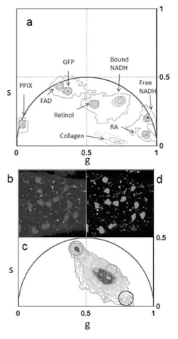
Figure 1 illustrates an example of phasor analysis of FLIM tissue images to identify tissue components. Figure 1a shows the phasor locations of pure chemical species for green fluorescent protein (GFP) in Tris buffer, Retinol in DMSO (pH 8.5), Retinoic acid in DMSO (pH 8.5), FAD in water (pH 7.4), free NADH in Mops buffer (pH 7), bound NADH in Mops buffer (pH 7) and lactate dehydrogenase, Protoporphyrin IX in dimethylformamide:methanol (pH 7). Figure 1b is the fluorescent intensity image of a semininiferous tubule from a mouse expressing GFP from an Oct4 transgene. Figure 1c is the phasor plot of the FLIM image acquired in Figure 1b. Figure 1d is the phasor color map, where the color in the phasor color map correspond to the color of the cluster of the phasor plot shown in Figure 1c.
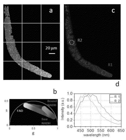
Figure 2.
The inventors have demonstrated the ability to identify the metabolic states of germ cells during differentiation using the phasor approach to FLIM. Figure 2a shows metabolite gradients in C. Elegans Germ line when excited at 740 nm. Figure 2b shows phasor plot selection using linear cluster that represents all possible relative concentrations of pure FAD (red), free NADH (blue), and NADH bound to MDH (green). Figure 2c shows the spectral image of the C. Elegans excited at 740 nm in the same field of view with R1 and R2 indicating regions of interest. The emission spectra R1 and R2 is shown in Figure 2d. The blue shift of the spectrum indicates an increase in bound NADH with respect to free NADH during differentiation.
This method allows for straightforward interpretation of intrinsic fluorescence signal from living tissues. Images of molecule species are obtained through their finger prints without the need to resolve and assign exponential components to the fluorescence species.
Suggested uses
This invention can be used to monitor metabolic states of cells, in particular stem cells, both in vitro and in vivo, and study cell fates. This invention can also be used to differentiate tissue composition.
Advantages
The invention uses the phasor approach for FLIM. This is a label-free method that allows for identification and quantification of relative concentrations of molecular species in living tissues.
Patent Status
| Country | Type | Number | Dated | Case |
| United States Of America | Issued Patent | 10,222,335 | 03/05/2019 | 2010-805 |
Contact
- Alvin Viray
- aviray@uci.edu
- tel: View Phone Number.