Variable Exposure Portable Perfusion Monitor
Patent Status
| Country | Type | Number | Dated | Case |
| United States Of America | Issued Patent | 11,710,236 | 07/25/2023 | 2022-890 |
Full Description
Background
Analyzing tissue perfusion is a common task in many medical procedures as it elucidates the viability of the tissue being examined. Monitoring perfusion parameters is of vital importance in wound healing, identifying ischemic occlusions, assessing success in reconstructive flap surgery and evaluating microcirculation impairment in patients with peripheral artery disease. Laser Speckle Imaging (LSI) is an established imaging modality that allows monitoring of perfusion in the tissue sample. Currently available systems are:
- Too bulky
- Very expensive (> $50,000)
- Require considerable energy and software/systems processing capacity
- Provide qualitative information only
For usage during surgery or post-operative monitoring, portable devices are preferred.
Invention
Inventors led by Prof. Guillermo Aguilar and Dr. Aditya Pandya at UCR have developed a novel, portable perfusion monitor by leveraging the computing capabilities of commercial vision processing systems-on-modules (SOMs) to perform Laser Speckle Imaging at video rates. The developed prototype acquires images to visualize variations in perfusion.

LSI imager prototype. An LSI camera was attached to the computer chip.
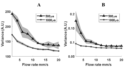
Average variance within ROI plotted for temporal (A) and spatial images(B) for two exposure times 500µs and 1000µs. Shaded regions represent +/- 1 standard deviation.
Advantages
The significant aspects of this novel device are:
Suggested uses
Applications that could benefit from this inexpensive, hand-held LSI device invention are:
State Of Development
The inventors have built a working prototype.
They are actively pursuing collaborators for optimization of the device and for testing with mice.
Inventions by Dr. Aguilar
Please review all inventions by Dr. Aguilar and his team at UCR
Contact
- Venkata S. Krishnamurty
- venkata.krishnamurty@ucr.edu
- tel: View Phone Number.

Other Information
Keywords
perfusion monitor, blood flow imaging, plastic surgery, reconstructive surgery, point of care, tissue imaging, tissue analysis