Dual Reflectance-Fluorescence Guided Surgical System
Background
Current real-time surgical imaging systems typically work by either direct visualization utilizing certain dyes and filters so normal tissue is seen as one reflected color and cancerous tissue as a visual fluorescent color; or a two camera system which requires the surgeon to alternate between viewing between reflected and fluorescent images displayed on two separate screens or switch between the images on a single monitor.
Technology Description
The UCSD research team of Drs. Roger Tsien, Quyen Nguyen and Paul Steinbach have developed a surgical imaging system that offers simultaneous viewing of reflected and fluorescent images, with complete channel separation and minimal time lags. The system can use a wide variety of fluorophores as well as fluorescence resonance energy transfer (FRET) probes and can be adapted to binocular vision systems. The fluorescent images provide for better localization of tumor margins for more precise excision, locating adjacent or involved nerves to avoid accidental severing; location of pre-cancerous tissue, arterial plaques. Methods have been developed and tested for integrating reflectance and fluorescence images; visualization of two simultaneous fluorphores (such as for visualization of tumor and nerve tissue) and three methods for utilizing FRET probes.
State Of Development
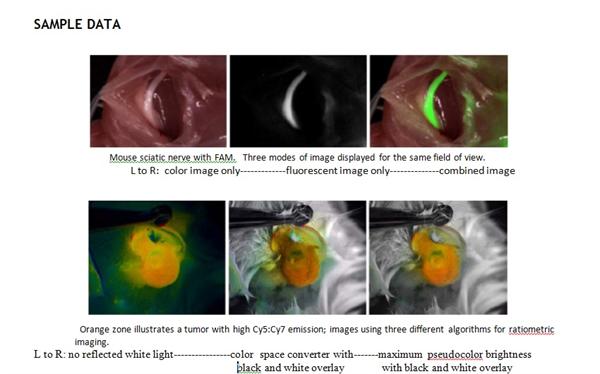
A prototype system has been assembled from conventional components consisting of an LED light source, lens, camera and computer and tested in small animal surgeries, including excision of tumor allografts and ratiometric imaging of tumor xenografts in a mouse breast cancer model using three different algorithms. A provisional patent application has been filed.
Intellectual Property Info
A provisional patent application has been filed.
Related Materials
- Fluorescence imaging in surgery. Orosco RK, Tsien RY, Nguyen QT. IEEE Rev. Biomed Eng. 2013; 6: 178-87 PMID 23335674 - 01/15/2013
- Real time in vivo molecular detection of primary tumors and metastases with ratiometric activatable cell penetrating peptides. Savariar EN et al. Cancer Res. 2013 Jan 15; 73(2): 855-64. PMID 23188503 - 01/15/2013
Patent Status
| Country | Type | Number | Dated | Case |
| United States Of America | Issued Patent | 10,231,626 | 03/19/2019 | 2013-185 |
| United States Of America | Published Application | 20190175021 | 06/13/2019 | 2013-185 |
Contact
- University of California, San Diego Office of Innovation and Commercialization
- innovation@ucsd.edu
- tel: View Phone Number.

Inventors
- Tsien, Roger Y.
Other Information
Keywords
surgical imaging, fluorescent, fluorescent resonance energy transfer, dyes, tumor, xenografts, cancer, breast,
Additional Technologies by these Inventors
- Personalized Protease fingerprinting for early cancer diagnosis
- Molecules for Labeling Peripheral Nerves for use in Image Guided Surgery and Other Clinical Applications
- Proteins that Efficiently Generate Singlet Oxygen Background
- Proteins That Fluoresce At Infrared Wavelengths Or Singlet Oxygen Upon Illumination