Molecules for Labeling Peripheral Nerves for use in Image Guided Surgery and Other Clinical Applications
Background
Surgical procedures often involve working close to vital nerve tissue and careful dissection is needed in order to preserve as much function as possible. Although some nerves are easily visualized, there are many types of procedures, such as the excision of tumors, the presence of inflammation or infection at the surgical site, scar tissue resulting from previous procedures or nerves encased in bone, where the precise location is not easily determined.
Currently nerves are typically visualized one at a time using fluorescent dyes that label nerves in either an antero- or retro-grade manner. These techniques have several drawbacks including excessively long labeling times to accumulate the imaging agent and limited accumulation of the agent along the axonal tract.
Technology Description
Researchers at UCSD have developed a fast, targeted approach to fluorescently label nerves that addresses all the disadvantages of current techniques. A targeting element, specific for neuronal tissue, limits dye accumulation in adjacent structures. Visualization of peripheral nerves occurs within a short time frame, enabling it for practical use during surgery, and improved accumulation of the imaging agent is obtained along the axonal tract of the nerve.
Advantages
• Systemic administration
• Visualization in real time at the surgical field
• Modular: so other dyes or molecules could be attached to targeting agent
• Specific accumulation in the axonal tract of neural tissue
• Potentially adaptable to delivery of neurotrophic factors to aid in repair and regeneration
State Of Development
• A patent application has been filed.
• A model system has been developed and tested in mice with specific and non-specific (control) targeting components
Results
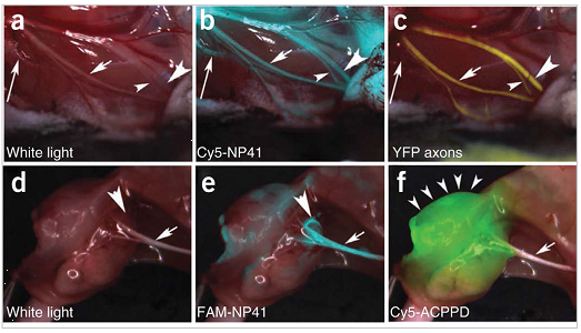
NP41 can highlight buried nerve branches invisible by standard illumination. (a–c) Right facial nerve and its arborizations in a thy1-YFP mouse treated with Cy5-NP41, viewed by (a) white light reflectance, (b) Cy5 fluorescence (pseudocolored cyan) overlaid on reflectance and (c) YFP fluorescence (pseudocolored yellow), also overlaid on reflectance. The short arrow marks a nerve branch visible by all three imaging modes. The arrowheads point to branches that are difficult to differentiate from muscle fascia in reflectance, but clearly distinguishable in both fluorescence images. The long arrow indicates a deeply buried branch visible only by Cy5-NP41 due to the better penetration of far-red wavelengths. (d–f) Left sciatic nerve (arrow) and its arborization in a mouse with a syngeneic 8119 mammary tumor graft1718, viewed by (d) white light reflectance, (e) FAM fluorescence 2 h after intravenous injection of NP41 (150 nmoles) (pseudocolored cyan, overlaid on reflectance) and (f) Cy5 fluorescence (pseudocolored green, overlaid on reflectance) from conjugates of activatable cell-penetrating peptides and dendrimers (ACPPDs). The large arrowheads in d and e point to a nerve branch buried under tumor, visible only by FAM fluorescence. Small arrowheads in f denote tumor.
Patent Status
| Country | Type | Number | Dated | Case |
| United States Of America | Issued Patent | 8,685,372 | 04/01/2014 | 2009-220 |
Contact
- University of California, San Diego Office of Innovation and Commercialization
- innovation@ucsd.edu
- tel: View Phone Number.

Inventors
- Tsien, Roger Y.
Other Information
Keywords
nerve tissue, visualization, fluorescent dyes, axonal tract,