Percutaneous Heart Valve Delivery System
Brief Description
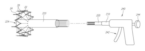
Researchers at University of California, Irvine have developed a novel percutaneous heart valve delivery system for coordinated delivery, positioning, repositioning, and/or percutaneous retrieval of percutaneously implanted heart valves. This system enables optimal placement of the transcatheter heart valve and may thereby significantly reduce the risk of paravalvular aortic regurgitation, myocardial infarction, or ischemia related to improper positioning.
Patent Status
| Country | Type | Number | Dated | Case |
| United States Of America | Issued Patent | 9,668,859 | 06/06/2017 | 2012-661 |
Technology Description
Transcatheter aortic valve replacement (TAVR) procedures require image-guidance during implantation to successfully deploy the heart valve into the correct position within the patient's aortic annulus. During the implantation procedure, only X-Ray can be used for image guidance. However, X-Ray is not sufficient for accurate visualization because it is a 2D projection of 3D anatomy that depends on the orientation angle of visualization. Currently, other imaging modalities such as CT, MRI, or ultrasound are used prior to the procedure and during the follow-up procedure, with the hopes that anatomical visualization can be directly correlated to the X-Ray images seen during the procedure. However, differences in contrast, resolution, and artifacts can produce differing results.
Correct valve positioning is crucial for treatment success and optimal outcomes after transcatheter valve implantation. However, most of the current technologies are limited by instant deployment, and once the valve is deployed, repositioning and/or percutaneous retrieval is not possible—or at least difficult or potentially problematic. Placement of the stented valve in a position that is too high (or proximal) can obstruct the coronary ostia in a case of aortic implantation, which may result in myocardial infarction or ischemia. Additionally, if the valve is placed too high in the aorta, it may embolize into the aorta, causing significant paravalvular regurgitation. On the other hand, implantation in a position that is too low (or distal) is accompanied by compression of the atrioventricular (AV) node in the membranous septum, which leads to conduction abnormalities.
Addressing this critical need for accurate placement of the transcatheter valve, researchers at University of California, Irvine have invented a percutaneous heart valve delivery system for coordinated delivery, positioning, repositioning, and/or percutaneous retrieval of the percutaneously implanted heart valves. This technology employs spring-loaded arms releasably engaged with a stent frame for controlling valve deployment and expansion. To control the state of deployment, this valve delivery system utilizes filaments passing through the distal end of a pusher sleeve in a self-expandable stent frame. This novel system, which also integrates a visualization system, allows optimal placement of the transcatheter heart valve, and may thereby significantly reduce the risk of paravalvular aortic regurgitation, myocardial infarction, or ischemia related to improper positioning.

Contact
- Alvin Viray
- aviray@uci.edu
- tel: View Phone Number.
Inventors
- Kheradvar, Arash
Other Information
Additional Technologies by these Inventors
- Percutaneous Heart Valve Delivery System Enabling Implanted Prosthetic Valve Fracture
- A distensible wire mesh for a cardiac sleeve
- Method to Improve the Accuracy of an Independently Acquired Flow Velocity Field Within a Chamber, Such as a Heart Chamber
- Growth-Accomodating Transcatheter Pulmonary Valve System
- System for Transcatheter Grabbing and Securing the Native Mitral Valve’s Leaflet to a Prosthesis
- Real-time 3D Image Processing Platform for Visualizing Blood Flow Dynamics
- Method for Synchronizing a Pulsatile Cardiac Assist Device with the Heart
- Fully Automated Multi-Organ Segmentation From Medical Imaging
- Simple, User-friendly Irrigator Device for Cleaning the Upper Aerodigestive Tract and Neighboring Areas
- Automated 3D Reconstruction of the Cardiac Chambers From MRI of Ultrasound
- Minimally Invasive Percutaneous Delivery System for a Whole-Heart Assist Device
