High External-Efficiency Nanofocusing for Lens-Free Near-Field Optical Microscopy
Background
The demand for imaging at the nanoscale has led to the invention and development of near-field scanning optical microscopy (NSOM). However, near-field scanning optical microscopy probes are inefficient in nanofocusing. Nanofocusing controls the concentration of light on nanometre scales, and a lack of control leads to blurred images. This prevents high-resolution images with valuable spatial information from being easily accessible at the nanoscale. Hardware and methods to increase control of nanofocusing would enhance imaging in NSOM.
Brief Description
Profs. Ruoxue Yan, Ming Liu, and their colleagues from the University of California, Riverside have developed a two-step sequential broadband nanofocusing technique with an external nanofocusing efficiency of ~50% over nearly all the visible range on a fibre-coupled nanowire scanning probe. By integrating this with a basic portable scanning tunneling microscope, the technology captured images with spatial resolution as low as one nanometer at high resolution. The high performance and vast versatility offered by this fibre-based nanofocusing technique allows for the easy incorporation of nano-optical microscopy into various existing measurement platforms.

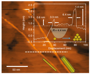
Fig. 1: High-resolution NSOM mapping. a, scanning tunnelling microscope topographic image of single wall carbon nanotubes on a gold film. Top inset: cross-sectional profile along the dashed line. Bottom inset: the possible configurations of the bundle.
Suggested uses
This new technique may be used:
- to measure stress levels of silicon for semiconductor chip manufacturers
- to dope field effect transistors to increase their efficiency
- for integration with cryo-chambers
- for applications in cryo-FIB (focused ion beam), cryo-FIB-SEM, or cryo-FIB-TEM for biological experiments
- to analyze PN junctions in solar cells
Related Materials
Patent Status
| Country | Type | Number | Dated | Case |
| United States Of America | Issued Patent | 11,054,440 | 07/06/2021 | 2019-116 |
Contact
- Venkata S. Krishnamurty
- venkata.krishnamurty@ucr.edu
- tel: View Phone Number.
Other Information
Keywords
near-field optical microscopy, lens-free, high external-efficiency, localized surface plasmon, focused ion beam, nanoscopy
