Volumetric Optical Coherence Tomography using a Passively Scanned Probe
Abstract
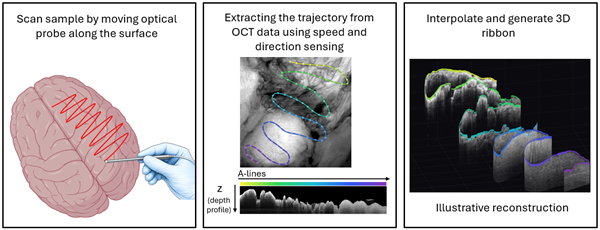
This technology introduces systems and methods for volumetric optical coherence tomography (OCT) using a handheld probe that passively scans the targeted tissue. Compared to active scanning OCT this allows a larger and more flexible field of view and eliminates the need for active scanning element. Unlike conventional passive OCT probes that only produce 2D cross-sectional images, this innovation reconstructs high-quality 3D volumetric images by assembling A-line data based on speckle decorrelation. This approach allows capturing high resolution volumes without needing any external motion-tracking systems, providing clinicians with detailed spatial information about tissue depth and lateral extent.
Full Description
Optical coherence tomography has entered the clinical domain with great fanfare seeing uptake in ophthalmology and cardiology. Both are applications that involve mechanical scanning. Aside from these, it has not deeply penetrated clinical scenarios where scanning is needed in or around the body. What is limiting uptake is scanning, which is both expense and complex. Passively scanned probes exist but they can only sense the speed of probe’s motion but not the direction, limiting OCT’s usefulness for 3D structure. External tracking can be used but it requires cameras, specialized probes, and is subject to resolution limits.
Researchers at the University of California, Davis have developed a technology that enables volumetric imaging with passive scanning OCT probes using only data contained in the collected signal, facilitating 3D tissue visualization. This dramatically increases the range of applications for OCT probes and lowers the cost of probes capable of 3D scanning, adding only minimal system complexity and requiring no external tracking hardware. (Reference figures at bottom.)
Applications
- Probe based scanning of 3D structures which contain structural information and can use optical coherence tomography
- Point‑of‑care diagnostics.
- Clinical tissue assessment for pathology diagnosis and monitoring.
- Dermatology and oncology for non-invasive lesion delineation.
- Surgical guidance and intraoperative imaging.
- Research settings require detailed volumetric tissue analysis.
- Industrial inspection systems for surface or material assessment
Features/Benefits
- Enables true volumetric (3D) imaging using passive scanning probes.
- Eliminates reliance on active/mechanical scanning hardware for 3D acquisition.
- Expands the field of view without requiring external motion-tracking systems.
- Allows 3D imaging of complex structures / scanning fields.
- Enables reconstruction of high-resolution volumes by leveraging speckle decorrelation to correct motion-related distortion.
- Enhances clinical usability by supporting a handheld, point-of-care workflow.
- Overcomes the inability of passive OCT probes to produce volumetric (3D) datasets.
- Removes dependence on mechanical scanners or external tracking to achieve 3D imaging.
- Reduces geometric distortion caused by nonuniform or irregular scan motion.
- Allows very compact and cost-effective probe design
Patent Status
Patent Pending
Other Information
Contact
- Michael M. Mueller
- mmmueller@ucdavis.edu
- tel: View Phone Number.

Inventors
- Kristen, Clara
- Marcu, Laura
- Thrapp, Andrew
Other Information
Keywords
3D imaging, a-lines, decorrelation, motion estimation, optical coherence tomography, passively scanned probe, speckle, volumetric imaging, handheld devices, field of view
Additional Technologies by these Inventors
- Fabrication Method for Side Viewing Miniature Optical Elements with Free-Form Surface Geometry
- Real-Time Fluorescence Lifetime Tracking
- Motor Drive Unit for Combined Optical Coherence Tomography and Fluorescence Lifetime Imaging of Intraluminal Structures
- Interferometric Near Infrared Spectroscopy and Fluorescence Lifetime Imaging
- Broadband Optical Rotary Junction with High Return Loss for Analysis of Plaques

